Animal Cell Diagram Labeled Flagella, Basics Of Animal Cell Biology Lovetoknow
- Pin By Bernadette Collins On Science Animal Cell Cell Structure Human Cell Structure
- Animal Cells Animal Cell Plant And Animal Cells Cell Model
- Animal Cell Worksheet Free Printable
- Cellworksheetkey
- Eukaryotic Cells Definition Parts Examples And Structure
- Plant Cell Simple English Wikipedia The Free Encyclopedia
- Cell Organisms And Their Functions
- Plant Cell Definition Labeled Diagram Structure Parts Organelles
- Lab Manual Exercise 1a
- Cell Biology Accessscience From Mcgraw Hill Education
Find, Read, And Discover Animal Cell Diagram Labeled Flagella, Such Us:
- Animal Cell Labeled Biologycorner Com Google Search In 2020 Animal Cells Worksheet Cells Worksheet Plant And Animal Cells
- Animal Cells Animal Cell Plant And Animal Cells Cell Model
- Animal Cells And The Membrane Bound Nucleus
- Shutterstock Puzzlepix
- Pearson The Biology Place
If you re searching for Charles By Shirley Jackson Plot Diagram you've reached the perfect location. We ve got 103 images about charles by shirley jackson plot diagram adding pictures, pictures, photos, wallpapers, and more. In such webpage, we also provide variety of graphics available. Such as png, jpg, animated gifs, pic art, symbol, blackandwhite, transparent, etc.
Cilia and flagella are also components of an animal cell and are crucial to the movement of the individual organisms of a cell.

Charles by shirley jackson plot diagram. The endoplasmic reticulum is a network of sacs that work to move chemic compounds throughout the cell. Learn more about the structures of animal cell anatomy using these hands on diagrams of an animal cell that we have collected for you in high quality. In these 101 diagramss the detailed structures of animal cell anatomy are illustrated in a high quality pictures.
Centrioles are about 500nm long and 200nm in width that are found close to the nucleus and helps in cell division. A flagellum f l e d l em. Cilia are smaller 5 20 um but are numerous.
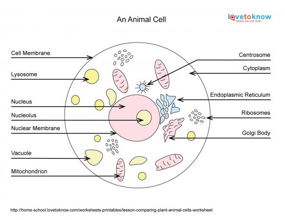
The animal cell diagram is widely asked in class 10 and 12 examinations and is beneficial to understand the structure and functions of an animal. Flagella is a lash like appendage that protrudes from the cell body of certain bacteria and eukaryotic cells termed as flagellatesa flagellate can have one or several flagella. Label the of an animal cell word box lysosome nucleus mitochondria cell membrane cytoplasm nucleolus cilia golgi apparatus cytoskeleton secretory vesicle rough endoplasmic reticulum smooth endoplasmic reticulum.
All these diagrams are printable and you are also provided the worksheet or unlabeled version of the diagrams. Where prokaryotes are just bacteria and archaea eukaryotes are literally everything else. Only 1 4 flagella occur per cell eg many protists motile algae spermatozoa of animals bryophytes and pteridophytes choanocytes of sponges gastro dermal cells of coelenterates zoospores and gametes of thallophytes.
They work to move fluid throughout the cell or a group of cells. All animal cells are multicellular and eukaryotic cells. Cilia and flagella are tiny hair like projections from the cell made of microtubules and covered by the plasma membrane.
They are paired tube like organelle composed of a protein called tubulin. From amoebae to earthworms to mushrooms grass. Learn more about the other biology.
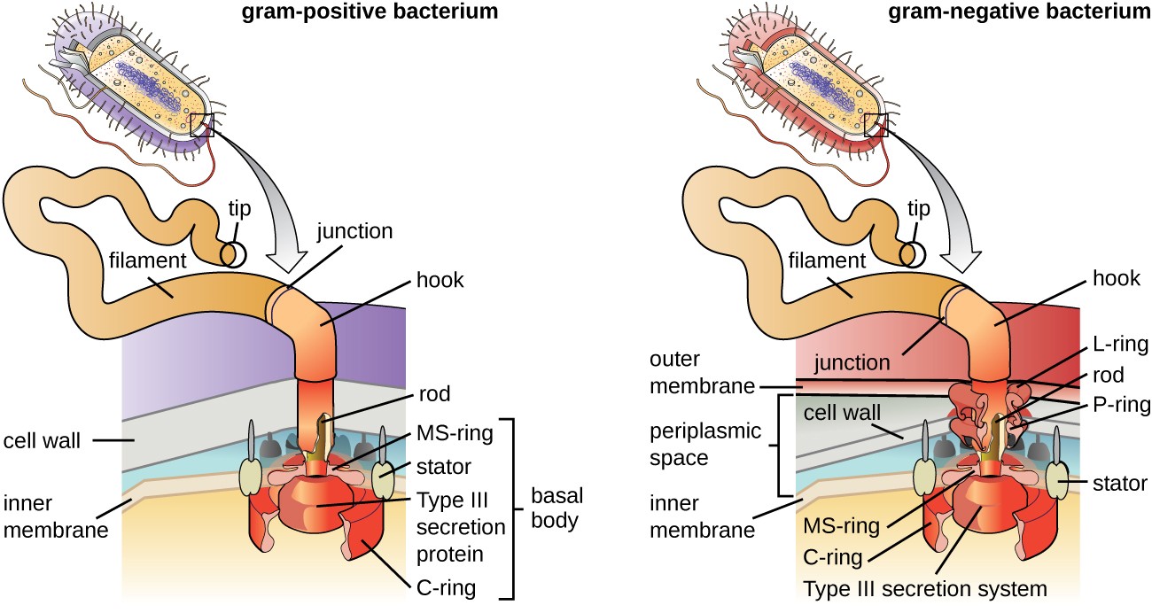
The primary function of a flagellum is that of locomotion but it also often functions as a sensory organelle being sensitive to chemicals and temperatures outside the. There are two types of cells prokaryotic and eucaryotic. The following animal cell diagram labeled.
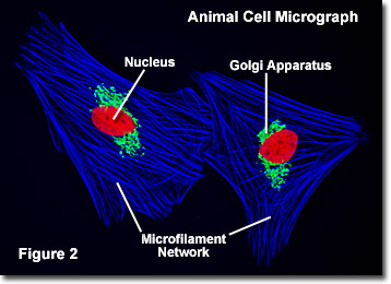
Flagella are longer 100 200 um but fewer. A brief explanation of the different parts of an animal cell along with a well labelled diagram is mentioned below for reference. Helping in cell division by allowing separation of chromosomes.
Cilia are hair like projections that have a 92 arrangement of microtubules with a radial pattern of 9 outer microtubule doublet that surrounds two singlet microtubules. Eukaryotic cells are larger more complex and have evolved more recently than prokaryotes. A labeled diagram of the animal cell and its organelles.
Charles By Shirley Jackson Plot Diagram, Unique Characteristics Of Prokaryotic Cells Microbiology
- 3 2 Unique Characteristics Of Eukaryotic Cells Geosciences Libretexts
- Shutterstock Puzzlepix
- Animal Cells Vs Plant Cells Science Cells Animal Cell Plant And Animal Cells
Charles By Shirley Jackson Plot Diagram, Migepaky
- Plant Cell Wikipedia
- Prokaryotic Cell Definition Examples Structure Biology Dictionary
- Cell Biology Accessscience From Mcgraw Hill Education
Charles By Shirley Jackson Plot Diagram, File Animal Cell Structure En Svg Wikimedia Commons
- Basic Cell Structures Review Article Khan Academy
- Structure Of Plant Cell Explained With Diagram
- 1
More From Charles By Shirley Jackson Plot Diagram
- Plant Cell Diagram Labeled 8th Grade
- Flowchart Programming Definition
- 2004 International 4300 Fuse Box Diagram
- Boron Electron Shell Diagram
- Pmos Circuit Diagram
Incoming Search Terms:
- Please Pick Up An Animal Cell Diagram And Start Labeling Ppt Download Pmos Circuit Diagram,
- Structure And Functions Of Cilia And Flagella Pmos Circuit Diagram,
- Lab Manual Exercise 1a Pmos Circuit Diagram,
- Pin On Micro Pmos Circuit Diagram,
- Https Www Sedelco Org Cms Lib02 Pa01001902 Centricity Domain 506 Bio 20cell 20structure 20and 20function 20chart 20and 20review Pdf Pmos Circuit Diagram,
- Animal Cell Model Diagram Project Parts Structure Labeled Coloring And Plant Cell Organelles Cake Animal Cell And Plant Cell Animal Cell Model Diagram Project Parts Structure Labeled Coloring And Plant Cell Organelles Pmos Circuit Diagram,


