Working Of Heart Diagram, 18 Heart Diagram Templates Sample Example Format Download Free Premium Templates
- 18 Heart Diagram Templates Sample Example Format Download Free Premium Templates
- Detailed Structure Of Frog S Heart
- Working Of A Human Heart Download Scientific Diagram
- Heart Anatomy Anatomy And Physiology
- Seer Training Structure Of The Heart
- Structure And Function Of The Heart
- Life Process 03 Working Of Heart Double Circulation 02 Class X Ntse Neet Foundation Youtube
- How Our Heart Works Structure And Function 3d Animation In English Youtube
- Heart Structure Function Facts Britannica
- 40 3a Structures Of The Heart Biology Libretexts
Find, Read, And Discover Working Of Heart Diagram, Such Us:
- Human Heart Anatomy Functions And Facts About Heart
- Sketch Of Human Heart Anatomy With Hand Written Labels Stock Illustration Download Image Now Istock
- How The Heart Works Heart Foundation
- 40 3a Structures Of The Heart Biology Libretexts
- Describe The Structure Of The Human Heart With The Help Of A Diagram Also Write A Note On Heart Beat From Science Life Processes Class 10 Meghalaya Board
If you re searching for Venn Diagram Probability Union you've reached the right place. We ve got 104 images about venn diagram probability union adding pictures, photos, photographs, backgrounds, and much more. In such web page, we also have variety of images out there. Such as png, jpg, animated gifs, pic art, symbol, black and white, transparent, etc.
The heart valves work the same way as one way valves in the plumbing of your home.

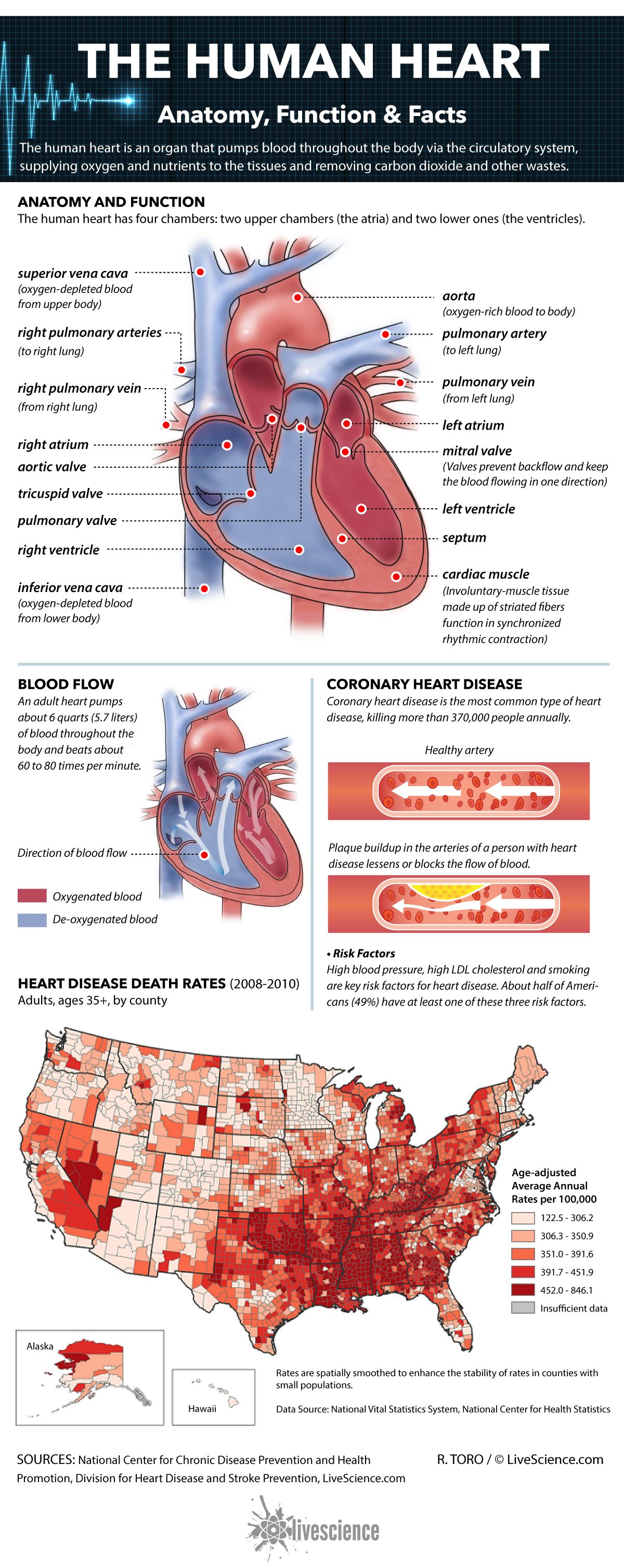
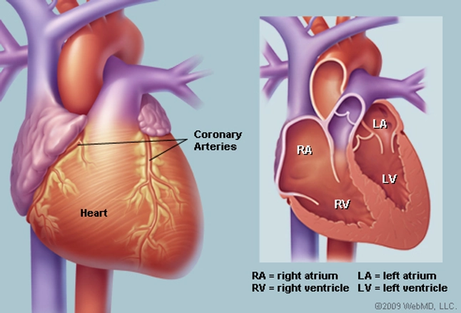
Venn diagram probability union. It is composed of four chambers many large arteries and many veins. The wall of the heart has three different layers such as the myocardium the epicardium and the endocardium. Heart diagram parts location and size location and size of the heart the heart is located under the rib cage 23 of it is to the left of your breastbone sternum and between your lungs and above the diaphragm.
Explain the working of human heart with a labeled diagram. The four chambers are called atrium and ventricles. The atrium and the ventricle of each side are separated by the atrioventricular septum.
The systemic circulation carries blood from the heart to all the other parts of the body and back again. A heart diagram labeled will provide plenty of information about the structure of your heart including the wall of your heart. They prevent blood from flowing in the wrong direction.
The heart is divided into four chambers two upper atria and two lower ventricles. The pulmonary circulation is a short loop from the heart to the lungs and back again. Each valve has a set of flaps called leaflets or cusps.
Lets check out heart diagram which can help you to understand functioning of the heart in a better way. Dear student the heart lies in the chest cavity between the lungs. Heart pumps pure blood to different parts of the body and then takes the deoxygenated blood from all the parts to the lungs for oxygenation.
It splits into two main branches and brings blood from. Heart diagram is the outlook figure of heart. Diagram of circulatory system working of circulatory system.
The heart is a muscular organ about the size of a closed fist that functions as the bodys circulatory pump. Heres more about these three layers. In the human heart diagram two atria and ventricles are separated from each other by a muscle wall called septum.
It takes in deoxygenated blood through the veins and delivers it to the lungs for oxygenation before pumping it into the various arteries which provide oxygen and nutrients to body tissues by transporting the blood throughout the body. Its major function is pumping blood continually muscular wall beat or contraction and blood pumping to all the parts of the body. The heart pumps blood through the network of arteries and veins called the.
Exterior of the human heart. Normally in a minute the heart beats 72 times and pumps around 1500 to 2000 gallons of blood per day. The human heart is situated under the ribcage.
The heart is a muscular organ about the size of a fist located just behind and slightly left of the breastbone. The septum separates the ventricles from each other and can be seen in the labeled heart diagram. The heart is about the size of a closed fist weighs about 105 ounces and is somewhat cone shaped.
Venn Diagram Probability Union, Working Of Human Heart Function Structure Of Heart Youtube
- How The Heart Works Diagram Anatomy Blood Flow
- Https Encrypted Tbn0 Gstatic Com Images Q Tbn And9gcqh1qywywrtrsuzo K6aqlxxoem4bee3bydstdtxvelneuwbkbl Usqp Cau
- How Our Heart Works Structure And Function 3d Animation In English Youtube
Venn Diagram Probability Union, Cardiovascular System Anatomy
- How To Draw The Internal Structure Of The Heart With Pictures
- How Does The Heart Work Science Made Simple
- Heart Anatomy Anatomy And Physiology
Venn Diagram Probability Union, Differences Between Working Ejecting Heart And Biventricular Working Heart
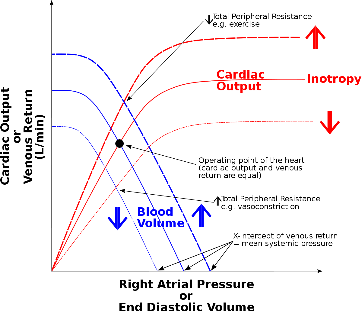
- Cardiac Output During 30 Minutes Of Working Heart Reperfusion Of 12 Download Scientific Diagram
- Draw And Lable Diagram Of Human Heart And Explain Its Working Brainly In
- The Heart Of The Matter National Geographic Society
More From Venn Diagram Probability Union
- Ba5412 Amplifier Circuit Diagram
- Allen Bradley E300 Wiring Diagram
- 3 Way Light Switch Wiring Diagram Multiple Lights
- 2013 Vw Tiguan Fuse Diagram
- Electron Configuration Orbital Filling Diagram Ws
Incoming Search Terms:
- Detailed Structure Of Frog S Heart Electron Configuration Orbital Filling Diagram Ws,
- Innovative Procedures To Get Your Heart Working Again Summa Health Vitality Electron Configuration Orbital Filling Diagram Ws,
- Describe The Working And Function Of Human Heart Briefly Long Question Carring 10 Marks Biology Topperlearning Com Gb2rjj66 Electron Configuration Orbital Filling Diagram Ws,
- Heart Diagram Coloring Sheet Awesome Anatomy Human Skeleton Coloring Human Heart Coloring In 2020 Anatomy Coloring Book Coloring Pages Coloring Books Electron Configuration Orbital Filling Diagram Ws,
- Effects Of Lactate Dehydrogenase Muscle Subunit M Ldh On The Heart Download Scientific Diagram Electron Configuration Orbital Filling Diagram Ws,
- How The Normal Heart Works Children S Hospital Of Philadelphia Electron Configuration Orbital Filling Diagram Ws,


