Microscope Cheek Cell Diagram, Images
- Cells Microscope Activity Unit Microscope Activity Science Cells Teaching Science
- Preparing Onion Cell Microscope Slide Investigation Instruction Sheet
- Hands On Activity Human Cheek Cells Science Supply
- Aqa Gcse 9 1 Biology For Combined Science Trilogy By Collins Issuu
- Cell Structure Microscope Lab By Buynomials Teachers Pay Teachers
- Microscope Lab
- Physiological Psychology
- How These 26 Things Look Like Under The Microscope With Diagrams
- Laboratory Microscopes Cells Fog Ccsf Edu
- Can You Expect To See Mitochondria While Using A Light Compound Microscope Socratic
Find, Read, And Discover Microscope Cheek Cell Diagram, Such Us:
- Images
- Virtual Microscope Cheek Cells Ppt Download
- Temporary Mount Of Human Cheek Cells Youtube
- Lab Manual Exercise 1
- Preparing Cheek Cell Microscope Slide Investigation Instruction Sheet
If you re looking for Ark Trailer Plug Wiring Diagram you've reached the perfect location. We ve got 104 graphics about ark trailer plug wiring diagram adding images, pictures, photos, backgrounds, and much more. In these webpage, we also provide variety of images available. Such as png, jpg, animated gifs, pic art, logo, blackandwhite, translucent, etc.
This cell do not have plastids vacuoles or cell wall.
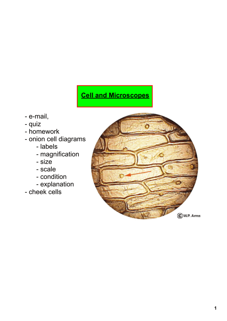
Ark trailer plug wiring diagram. Robert hooke 1650 also famous for his law of elasticity in physics observed and drew cells using a compound microscope. In this figure cheek cells stained with methylene blue. An onion is a multicellular consisting of many cells plant organismas in all plant cells the cell of an onion peel consists of a cell wall cell membrane cytoplasm nucleus and a large vacuole.
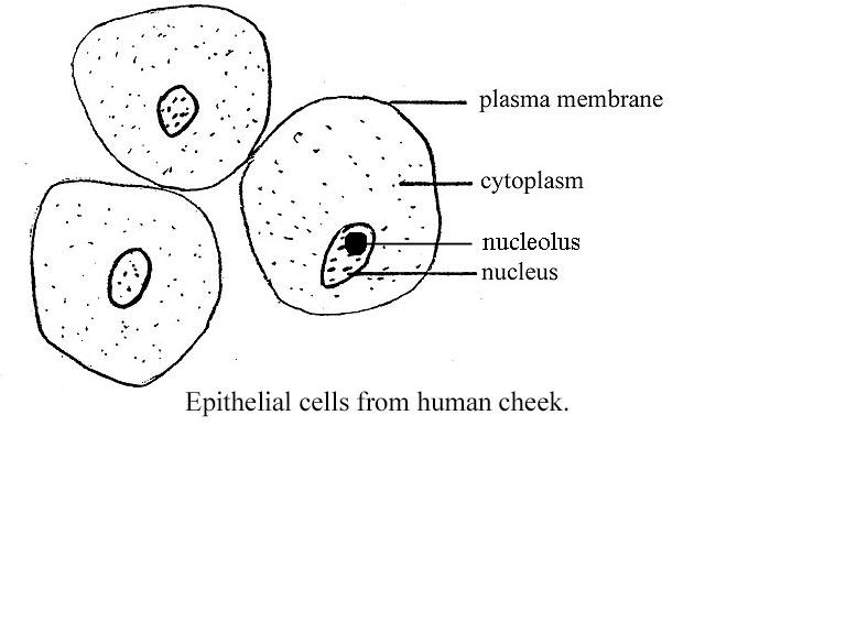
Image result for human cheek cell diagram chamber of secrets unbiol1 histology lab with answers cell structure and functions cbse science class 8 chapter wise. Before exploring the details of cell structure lets understand the differences in the structure of an onion cell and a human cheek cell. They have irregular cellular thin boundaries which contains jelly like cytoplasm and the cytoplasm are granular.
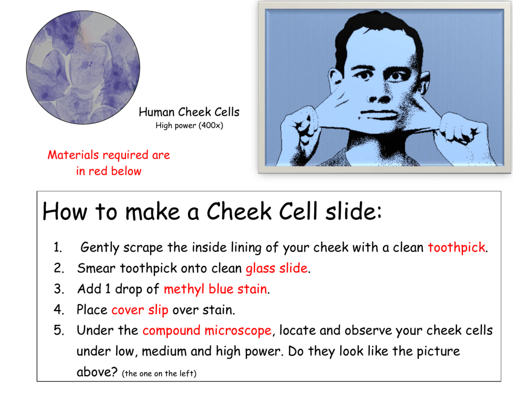
The nucleus at the central part of the cheek cell contains dna. Human cheek cells are made of simple squamous epithelial cells which are flat cells with a round visible nucleus that cover the inside lining of the cheekc. Add a single droplet of water squeezed from a plastic pipette onto the center of the slide.
Human cheek cells are observed under microscope 1. Cells are polygonal or flat in shape and structure 2. Introduction to the compound microscope cell structure function microscopy and cell diversity a labeled diagram of the plant cell and functions of its organelles.
Rotate the toothpick in the water to release the human cheek cells into the drop of water. These cells line the buccal cavity in humans and are usually shed during mastication and even talking. Cell structure aqa.
When a drop of methylene blue is introduced the nucleus is stained which makes it stand out and be clearly seen under the microscope. In addition you can learn the cell structure by looking at your own cheek cells through identifying cell membrane cytoplasm organelles and nucleus. Although the entire cell appears light blue in color the nucleus at the central part of the cell is much darker which allows it to be identified.
Cheek cells under the microscope. A replica of robert hookes compound. The cells in the cheeks are eukaryotic cells with a defined nucleus enclosed inside a nuclear membrane along with other cell organelles.
They are generally made up of squamous epithelium cells.
Ark Trailer Plug Wiring Diagram, Cell Lab Doc Plant And Animal Cells Microscope Lab Objectives Students Will Discover That Onions Are Made Up Of Cells Students Will Observe Onion Course Hero
- Polymath At Large The Little Things That Keep Us Going
- 2
- Difference Between Onion Cell And Human Cheek Cell Pediaa Com
Ark Trailer Plug Wiring Diagram, Virtual Microscope Cheek Cells Ppt Download
- Difference Between Onion Cell And Human Cheek Cell Pediaa Com
- Preparing Onion Cell Microscope Slide Investigation Instruction Sheet
- Lesson 2 Mount A Slide Look At Your Cheek Cells Rs Science
Ark Trailer Plug Wiring Diagram, Hands On Activity Human Cheek Cells Science Supply
- Onion And Cheek Cells By Dr Dave S Science Teachers Pay Teachers
- Solved 9 Imagine You Are Viewing A Specimen Directly Und Chegg Com
- Microscope Cell Lab Cheek Onion Zebrina Schoolworkhelper
More From Ark Trailer Plug Wiring Diagram
- Heart Pain Location Diagram
- Dicot Stem Diagram
- Buzzer Circuit Using Transistor
- Circuit Diagram Examples
- Calcium Atom Diagram
Incoming Search Terms:
- Histology Lab With Answers Calcium Atom Diagram,
- Cell Lab Doc Plant And Animal Cells Microscope Lab Objectives Students Will Discover That Onions Are Made Up Of Cells Students Will Observe Onion Course Hero Calcium Atom Diagram,
- Human Cheek Cell On Pcs A Bright Field Microscopy B Phase Download Scientific Diagram Calcium Atom Diagram,
- Learning Activity 1 1 Learning About Cells And Microscopes Calcium Atom Diagram,
- Images Of Human Epithelial Cheek Cells A Image Recorded By A Download Scientific Diagram Calcium Atom Diagram,
- How To Draw Human Cheek Cell Most Easy Way Step By Step Youtube Calcium Atom Diagram,


