Cell Wall Diagram Drawing, 2 4 1 Draw And Label A Diagram To Show The Structure Of Membranes Youtube
- Ib Biology Topic 2 4 1 Draw And Label The Plasma Membrane Youtube
- Making Biological Drawings Introduction To Biology
- Learning By Questions
- 2 3 Eukaryotic Cells Bioninja
- How To Draw A Plant Cell Plants Botany Easily Quickly Well Labelled Diagram Youtube
- Cell Wall Vector Images Stock Photos Vectors Shutterstock
- Https Encrypted Tbn0 Gstatic Com Images Q Tbn And9gcqp8y E5zo0x9vd91csfam9iawug Wicl L7g4r G3janczvvwe Usqp Cau
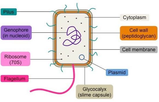
- Prokaryotic Cells Bioninja
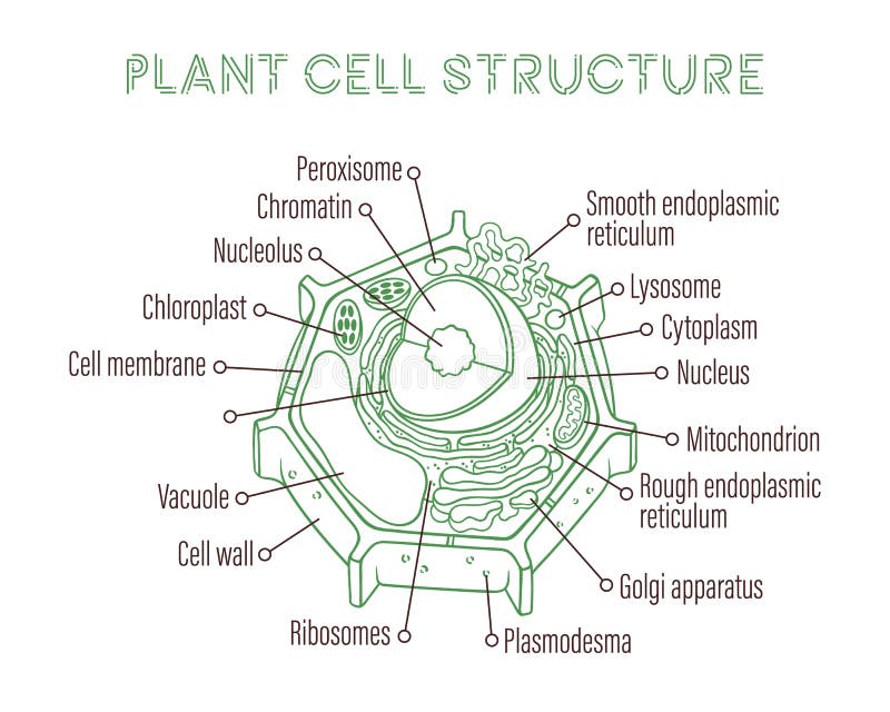
- Generalized Plant Cell
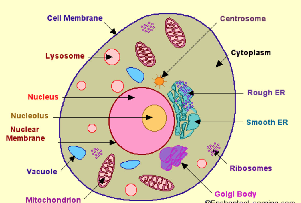
- Printable Animal Cell Diagram Labeled Unlabeled And Blank
Find, Read, And Discover Cell Wall Diagram Drawing, Such Us:
- Plant Cell Drawing With Labels Plant And Animal Cell Pictures With Labels In Cell Biological Cells Worksheet Animal Cell Plant Cells Worksheet
- Draw A Well Labelled Diagram Of Plant Cell Sarthaks Econnect Largest Online Education Community
- Schematic Drawing Of A Cylindrical Cell Wall Layer In A Global View Download Scientific Diagram
- Draw A Diagram Of A Plant Cell And Label At Least Eight Important Organelles In It
- Pin On 4eme Uaa3 Ultra Structure Cellulaire
If you are searching for Lewis Dot Structure Fluorine you've reached the right place. We ve got 104 images about lewis dot structure fluorine including images, photos, photographs, wallpapers, and more. In these page, we additionally provide variety of images available. Such as png, jpg, animated gifs, pic art, logo, blackandwhite, translucent, etc.
Then connect the.

Lewis dot structure fluorine. A plant cell may consist of either primary wall only or the cell may contain both primary and secondary walls fig. An additional non living layer present outside the cell membrane in some cells that provides structure protection and filtering mechanism to the cell is the cell wall. This is why its called the middle lamella even though it is the outermost layer.
Stock illustration by tussik 2 7 view of a sliced open blood vessel drawing by somersault1824 5 91 alveoli anatomy respiration drawing by megija 3 44 empty prison cell background stock illustration by backgroundstore 1 277 empty room white wall tile floor drawings by kostins 1 25 vector abstract mosaic design stock illustrations by. A cell wall is a rigid semi permeable protective layer in some cell types. This will also help you to draw the structure and diagram of cell wall in plants.
It is 10 25 nm in thickness. To represent these pores erase three or four small sections of each circle. The nucleus of an animal cell has pores in it called nuclear pores.
It gives shape to the cell. This outer covering is positioned next to the cell membrane plasma membrane in most plant cells fungi bacteria algae and some archaea. Draw two circles for the cell nucleus.
The nucleus is one of the larger structures of the cell. The first formed wall of a cell is the primary wall. Gross structure of cell wall.
It usually grows in surface area. Cell illustrator is a software tool that enables biologists to draw model elucidate and simulate complex biological processes and systems. A cell wall is multilayered.
Animal cells however do not have a cell wall. Jan 22 2018 step by step tutorials on drawing biology diagrams. In a plant cell the cell wall is made up of cellulose hemicellulose and proteins while in a fungal cell it is composed of chitin.
The cell wall has many important functions in a cell including protection structure and support. The single circular double stranded chromosome is the bacterial genome. That you can drag and drop so you dont have to spend time drawing each element.
Form the nucleus by drawing two circlesa larger circle that takes up around 10 of the cell with a slightly smaller circle inside it. Its an online app that contains a library of pre made cells proteins membrane shapes organs lab equipment etc. Lps teichoic acid etc surrounding the bacterium like a shell and lies external to the cytoplasmic membrane.
Lewis Dot Structure Fluorine, Draw A Diagram Of A Plant Cell And Label At Least Eight Important Organelles In It
- The Cell Wall Biology For Majors I
- Ib Biology Topic 2 4 1 Draw And Label The Plasma Membrane Youtube
- How To Draw Plant Cell For Class 9 To 12 Step By Step Demonstration Simple And Easy Bio Diag Youtube
Lewis Dot Structure Fluorine, Solved The Diagram Below Is A Student Drawing Of An Onion Chegg Com
- Prokaryotic Cells Bioninja
- Difference Between Plant And Animal Cells Cells As The Basic Units Of Life Siyavula
- Making Biological Drawings Introduction To Biology
Lewis Dot Structure Fluorine, The Cell More Information Cell Wall Atlas Of Plant And Animal Histology
- How Does The Cell Membrane In The Drawing Above Help To Maintain The Health Of This Cell A By Brainly Com
- Plant Cell Drawing With The Main Cell Compartments For Recombinant Download Scientific Diagram
- Ib Biology Notes 2 3 Eukaryotic Cells
More From Lewis Dot Structure Fluorine
- Function Of Heart Diagram
- The Rain Cycle Diagram
- Venn Diagram Numbers
- Diagram Of The Heart Oxygenated And Deoxygenated Blood
- Orbital Diagram For Nitrogen
Incoming Search Terms:
- Cellular Organelles And Structure Article Khan Academy Orbital Diagram For Nitrogen,
- The Fluid Mosaic Model Of The Cell Plasma Membrane For Dummies Cell Membrane Coloring Worksheet Plasma Membrane Cell Membrane Orbital Diagram For Nitrogen,
- Cell Structure Orbital Diagram For Nitrogen,
- 2 3 Eukaryotic Cells Bioninja Orbital Diagram For Nitrogen,
- Generalized Plant Cell Orbital Diagram For Nitrogen,
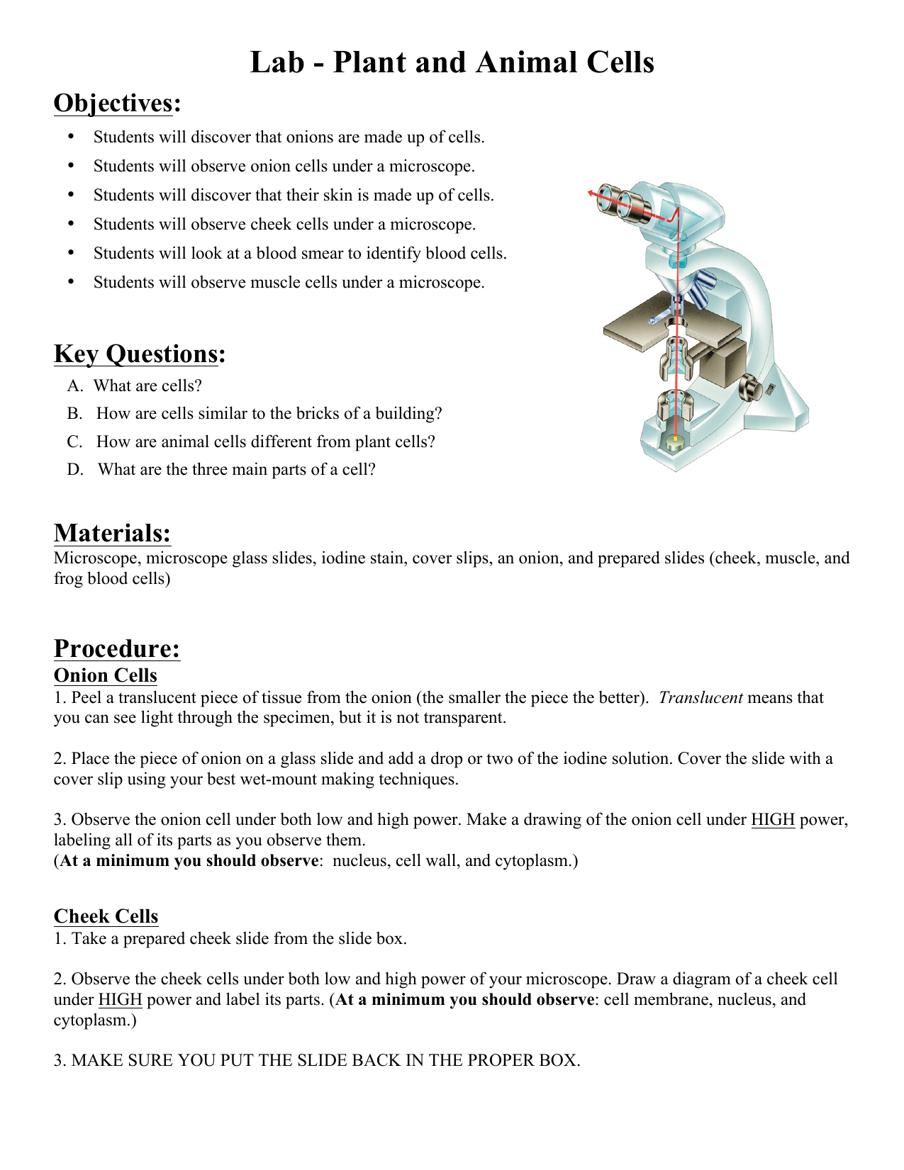
- 1 Lab Plant And Animal Cells Orbital Diagram For Nitrogen,


